All Products
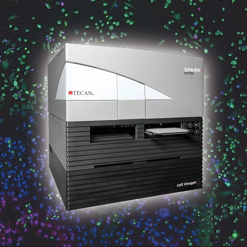
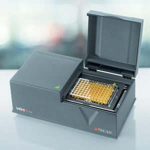
Spark® Cyto Multi-mode Plate Reader
PLATE READER WITH LIVE CELL IMAGING AND REAL-TIME CYTOMETRY
Spark Cyto is a multi-mode plate reader with fluorescence imaging and cytometry capabilities, unlocking new possibilities for your cell-based research. By combining live cell imaging with industry-leading detection technologies, you now have the ability to unite qualitative and quantitative information into unique multi-parameter data sets.
Your cells don’t stay static when you leave the lab, so your research requires a dynamic instrument that ensures you never miss a key event. Spark Cyto works in real -time, using parallel data acquisition and analysis- to deliver meaningful insights faster than before.
More insights delivered in real-time, uncover new approaches with Spark Cyto
More cells analyzed
Spark Cyto uniquely brings together top-of-the-range camera components with proprietary patent-pending technology to ensure that you can truly investigate your entire cell population. It gives you the ability to record the whole well area of a 96- or 384-well microplate with just one image, without tiling or distortion. This means that you never miss a cell when investigating the total cell population in a microplate well. Spark Cyto includes three magnification levels combined with four channels for fluorescence and bright field imaging, enabling high quality cell analysis for a wide variety of applications.
More parameters measured
Spark Cyto is available in configurations, building on the foundation of the Spark multimode reader platform. It combines sophisticated imaging with proven multimode reader capabilities, allowing you to define new approaches to your research and obtain robust orthogonal data faster than ever before.
More throughput for your experiments
Spark Cyto can be extended with an automated multi-plate cell incubator or integrated in Tecan’s fully automated workflow solutions.
More experimental control
Designed to handle a broad range of common cytometry applications, Spark Cyto gives you a new level of experimental control without compromising on ease of use and convenience. Five predefined methods for common cytometry applications offer a straightforward approach to image acquisition and analysis, complemented by additional features such as ‘user-defined’ parameters and Real Time Experimental Control (REC™), making it possible for you to unlock new application possibilities
Spark Cyto capabilities
Applications
- Nuclei counting
- Transfection efficiency
- Cell viability
- Apoptosis
- Confluence assessment
- Cell migration and wound healing
- ELISAs
- Low-volume DNA/RNA quantification
- Nucleic acid labeling efficiency
- Protein quantification
- Reporter gene assays
- HTRF®, DELFIA®, LanthaScreen®
- Transcreener®
- DLR®
- BRET – including NanoBRET®
Detection modes
- Fluorescence imaging (blue, green, red, far red)
- Bright field imaging
- Digital phase contrast-imaging
- Absorbance – incl. UV/VIS
- Fluorescence top and bottom
- Time resolved fluorescence (TRF)
- Full spectral scanning capability for all measurement modes
- FRET
- TR-FRET
- Fluorescence polarization (FP)
- Luminescence – glow, flash, multicolor, scanning
- AlphaScreen®, AlphaLISA® and AlphaPlex®
Additional features
- Reagent dispensers with heater and stirrer
- Humidity Cassette
- NanoQuant Plate™
- QC tools for IQ/OQ services
- Spark-Stack™ microplate stacker
- Multi-plate automated cell incubator extension
DOWNLOADABLE BROCHURE:
Spark® Cyto Multi-mode Plate Reader